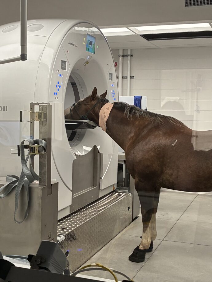
A 19-year-old quarter horse with a salivary stone in his cheek was the “model patient” for a veterinary hospital’s new CT scanner.
Joe Juice, who is a member of the teaching herd at the UC Davis Center for Equine Health (CEH) in the US, developed the hard mass just as the William R Pritchard Veterinary Medical Teaching Hospital’s (VMTH) diagnostic imaging service was preparing to open a new imaging centre. Part of that preparation included training sessions imaging animals on the new equipment, including a large-bore equine computed tomography (CT) scanner.
“Four years ago, Joe Juice had a sialolith removed from the right side of his face at the VMTH,” a UC Davis spokesperson said. “He recovered quickly and returned to his role as a member of the CEH teaching herd.
“Fast forward to the summer of 2025, when Joe Juice’s CEH care team discovered a visible bump on his cheek and suspected another sialolith based on his history.”
Sialolithiasis, or the formation of salivary stones, is uncommon in horses. The hard masses usually start small, as items such as small pieces of food make their way into the salivary duct and grow as layers of calcium form around them. As they grow, they can cause inflammation, infection and difficulty eating, as well as potentially altering saliva production and affecting digestion.
A study reported recurrence of sialoliths after surgical removal in 24% of cases, the average time to recur noted as just under three years.
“Since Joe Juice was already in need of imaging to confirm the sialolith, his veterinary team decided to utilise him as one of the first training models for the new equine CT scanner,” the spokesperson said.
The scan showed a large oval structure, of some 2x3x6cm in the soft tissue of Joe Juice’s cheek.
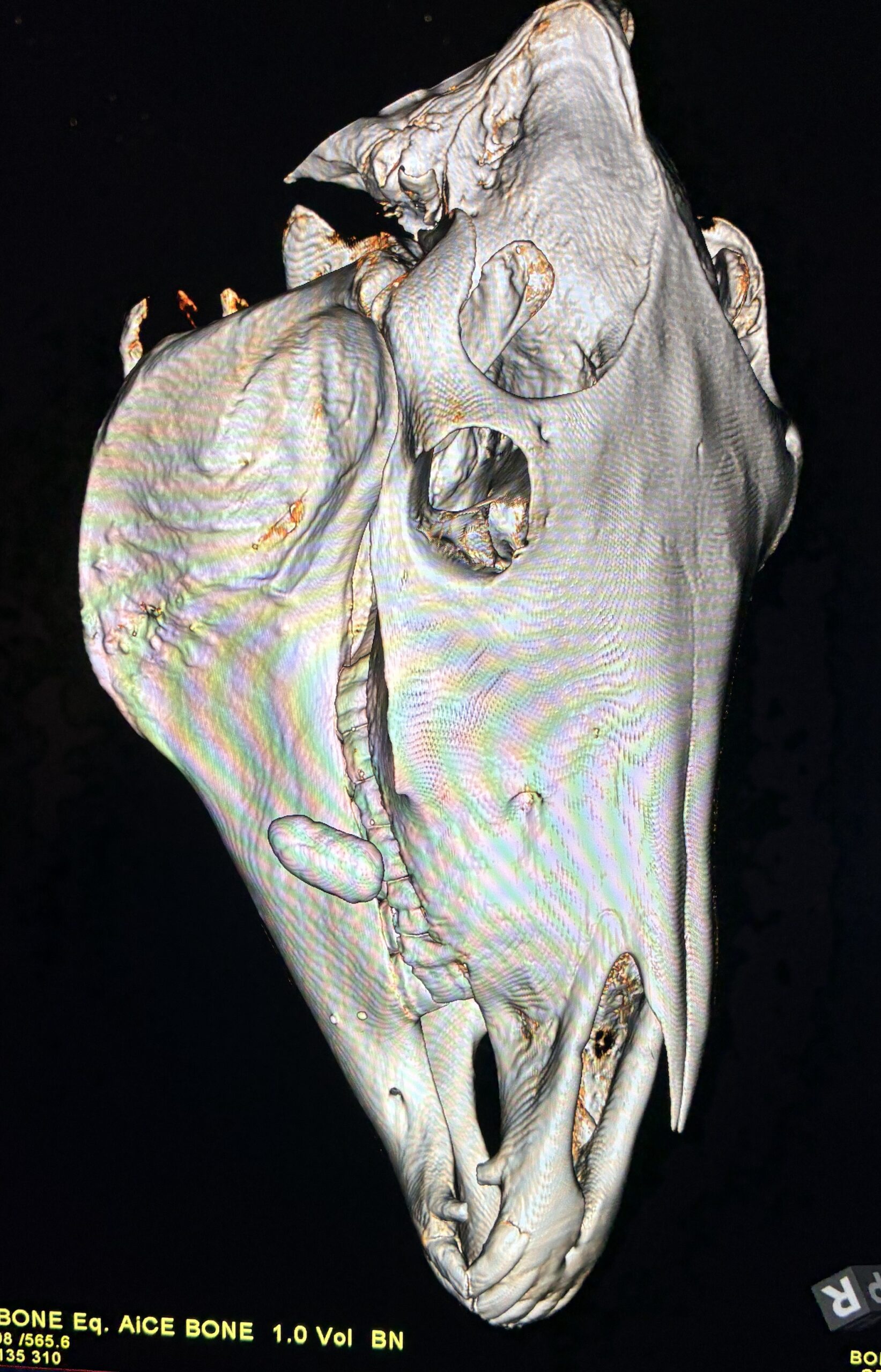
The stone can be seen on the left of the scan image
“Joe Juice was a model patient for our standing CT training,” said Mathieu Spriet, director of imaging services at the VMTH. “His sialolith was definitively diagnosed with the standing CT, giving surgeons a clear and concise assessment of the growth. This equipment’s ability to determine injuries and disease without full anesthesia is already proving to be tremendously valuable for our patients.”
The sialolith was removed, under standing sedation, by Scott Katzman, Stephanie Ortiz and David Orozco-Lopez, and Jo Juice was able to go home to the CEH shortly afterwards.
“Joe’s case is a great example of the importance of routine check-ups for your animals,” said Dr Katzman. “His first sialolith was discovered during a routine procedure several years ago, and this second one was discovered early because his care team routinely examines him for abnormalities. It’s also a great example of what we’re able to do with sedated standing procedures, instead of full anesthesia.”
The new equine CT scanner allows more diagnostic capabilities for horses and other large animals, the UC Davis spokesperson said, adding that the previous machine could only scan the limbs and heads of fully anesthetised horses. This one is integrated onto a platform that can be moved vertically and horizontally, so horses’ heads and limbs can be scanned under standing sedation, and it allows scanning of the stifles, pelvis and entire vertebral column.
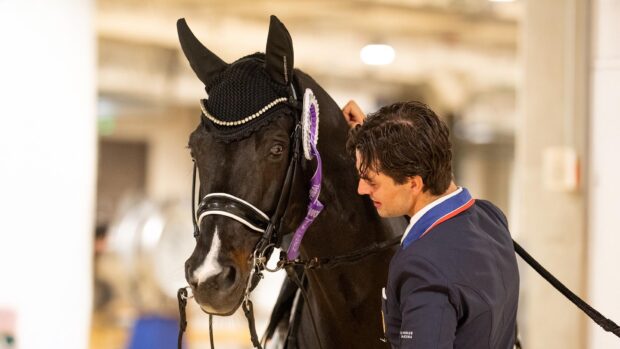
“We’re so grateful for the imaging, anesthesia, and surgery expertise at the VMTH,” said CEH director Carrie Finno. “Joe Juice has been a star member of our herd for 17 years. He is one of our go-to horses when it comes to teaching veterinary students. His kindness and patience help them learn about everything from lameness exams to cardiology.”
- To stay up to date with all the breaking news from major shows throughout 2025, subscribe to the Horse & Hound website
You may also be interested in:
New US dope-test rule after horses given euthanasia drug as ‘calmer’ for competitions

‘A frightening scene’: days-old foal found with broken jaw saved by expert vets

Examining the equine brain through CT scanning *H&H VIP*
X-ray may be the bread and butter of diagnostic imaging, but computed tomography (CT) goes one step further to allow

‘Horses will be horses’: blackthorn embedded in ligament remains undetected for a year

Subscribe to Horse & Hound magazine today – and enjoy unlimited website access all year round